PiSITE examples
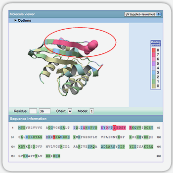
How to access each entry
Ras proteins are monomeric GTPases and control intracellular signaling cascades. Ras proteins regulate signal pathways involving actin cytoskeletal integrity, proliferation, differentiation, cell adhesion, apoptosis, and cell migration. Ras proteins are famous because they implicated in development of cancer.
PiSITE provides 11 different binding states, including 9 different binding partners for this protein. The Ras proteins are activated by Guanine nucleotide exchange factors (GEFs) and one of the well-known activator of Ras proteins is a Son of sevenless (SOS) protein. The second binding state provides the interaction between Ras and SOS protein.
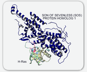
The part of interaction sites between Ras and SOS proteins are colored magenta. The color of each residue of a Ras protein shows the number of binding partners of each residue, and the residues contacting many partners are colored magenta. Interestingly, this magenta-colored region, including residues 32 to 42, corresponds to the effector region of a Ras protein. This region of a Ras protein is known as a hot spot and has been identified as a neutralizing epitope of hRas[1].
references
- Sigal, I.S., Gibbs, J.B., D'Alonzo, J.S. and Scolnick, E.M. (1986) Identification of effector residues and a neutralizing epitope of Ha-ras-encoded p21. Proc Natl Acad Sci USA, 83, 4725-4729.
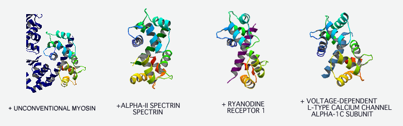
Calmodulin
Calmodulin is a calcium-binding protein found in all eukaryotic cells. This protein has two symmetrical domains, separated by a flexible hinge region. Calmodulin bind and regulate many different protein targets, involving many different cellular functions.
PiSITE provides 10 different binding states, including 8 different binding partners for this protein. Unfortunately, one of the most famous Calmodulin binding protein, Calmodulin-dependent-protein kinase II-alpha, can not found in binding partners because only the too short fragment of the protein is included in complexes. However, other famous calmodulin binding proteins, such as myosine light chain, can be found. In all binding states, calmodulin shows same binding style. Calmodulin wraps around the alpha helix of its binding partners by using the hinge region. Calmodulin tightly bind to a wide range of different target proteins by changing its global conformation.
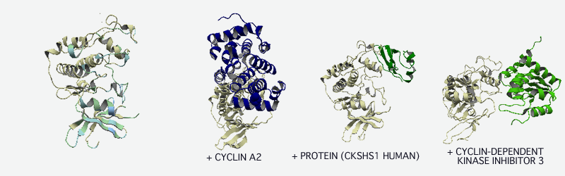
Cdk2
Cyclin-dependent kinases (CDK) belong to a group of protein kinases originally discovered as being involved in the regulation of the cell cycle. cyclin-dependent kinase is activated by association with a cyclin, forming a cyclin-dependent kinase complex.
PiSITE provides 8 different binding states, including 7 different binding partners, for this proteins. More than half of the binding states include cyclins ("CYCLIN A2" or "GAMMA HERPESVIRUS CYCLIN") as binding partners. In the 1st binding state, Cdk2 binds to "CKSHS1", and in the 5th binding state, Cdk2 binds to "CYCLIN-DEPENDENT KINASE INHIBITOR 3". In both cases, the binding sites do not correspond to that of cyclin. This can be easily checked from the picture colored by the number of binding partners.
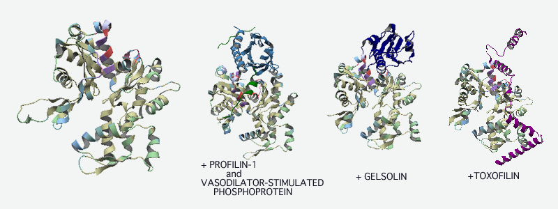
Actin
Actin is a globular protein found in all eukaryotic cells. It is the monomeric subunit of microfilaments, one of the three major components of the cytoskeleton, and of thin filaments. Actin participates in many important cellular functions, including muscle contraction, cell motility, cell division and vesicle movement.
PiSITE provides 13 different binding states, including 10 different binding partners, for this protein. As shown in the molecule viewer in default state, actin has a hot spot, including residues 340 to 360. Most of actin-regulating proteins, such as "PROFILIN", "GELSOLIN" and "TOXOFILIN" bind to this region. These binding proteins function in different ways. However, all of them have the function to inhibit the polymerizations of actin molecules.